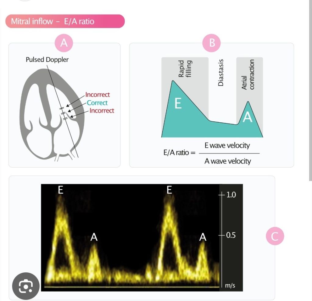
E/A
Pagi sabtu ni kita belajar diastology hahah.
Think of transmitral Doppler as watching blood move from the left atrium (LA) into the left ventricle (LV) during diastole.
The shape of the waveform tells us:
How well the LV relaxes
How stiff the LV is
Whether filling pressures are high
The basic story
After the heart squeezes:
The LV relaxes
LV pressure falls
When LV pressure becomes lower than LA pressure → the mitral valve opens
Blood flows into the LV
This filling creates the Doppler waves.
The 2 main waves
1. E wave = Early filling
This is the first wave.
Blood rushes passively from LA → LV because the LV is relaxing and “sucking” blood in.
Good relaxation → bigger E wave
Poor relaxation → smaller E wave
Easy memory:
E = Early passive Emptying into LV
2. A wave = Atrial contraction
This is the second wave.
The atrium squeezes at the end of diastole to push extra blood into the LV.
If LV is stiff, the atrium must push harder → bigger A wave
Easy memory:
A = Atrial kick
E/A ratio
This is the quickest way to look at diastolic function.
�
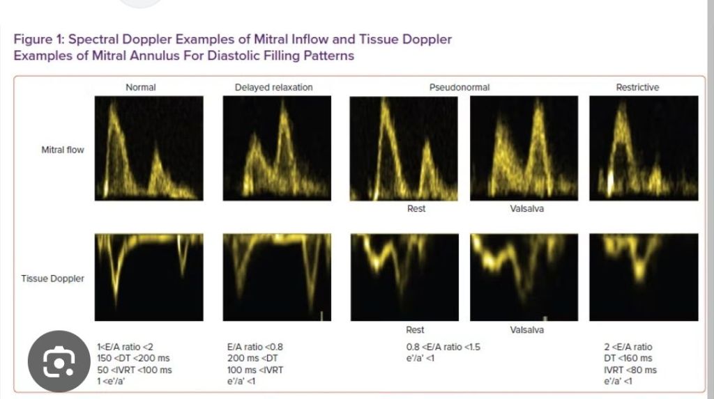
Normal young heart
E > A
Ratio usually >1
Because relaxation is good.
Impaired relaxation
LV relaxes slowly
Less early filling
Atrium must help more
So:
Small E
Big A
E/A <1
Typical pattern:
“Old stiff ventricle.”
Restrictive filling
LV is very stiff and filling pressure is high.
Blood rushes rapidly into LV early:
Very tall E
Small A
Short deceleration time
This is usually a severe pattern.
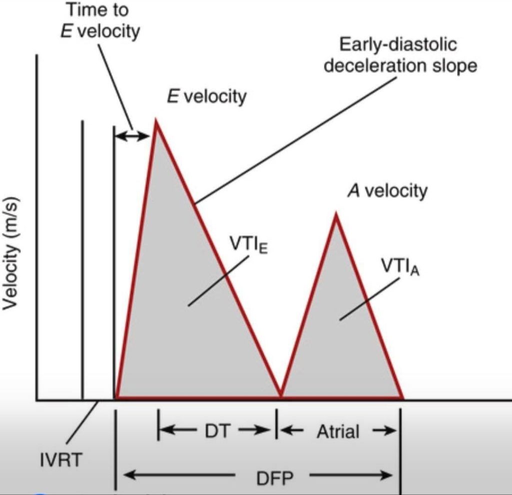
Deceleration Time (DT)
This is how fast the E wave falls back down.
Long DT
Flow slows gradually.
Usually means:
Slow relaxation
Less compliant ventricle
Short DT
Flow stops quickly because LV pressure rises rapidly.
Usually means:
High filling pressure
Restrictive physiology
IVRT (Isovolumic Relaxation Time)
Time between:
Aortic valve closing
Mitral valve opening
�
It reflects how fast the LV relaxes.
Long IVRT
LV relaxes slowly.
Short IVRT
LA pressure is high, so mitral valve opens earlier.
Pseudonormal pattern
Sometimes the waveform looks “normal”:
E/A around 1–1.5
But actually:
LV relaxation is abnormal
LA pressure is elevated
The high LA pressure artificially increases the E wave.
Pening…
That is why we also check:
Tissue Doppler (e′ velocity)
LA size
Pulmonary venous flow
Simple way to think about it
Transmitral Doppler is basically asking:
“How easily does blood enter the LV?”
Relaxed LV → smooth easy filling
Stiff LV → atrium struggles
High pressure LV → blood rushes in abnormally fast
So the waveform is really a conversation between:
LA pressure
LV relaxation
LV stiffness/compliance
One-line summary
E wave = passive filling
A wave = atrial push
E/A ratio + DT + IVRT = clues to LV relaxation and filling pressure


Leave a comment